Fig. 1
Crush injury of the distal tibia with loss of plafond and comminuted fibula
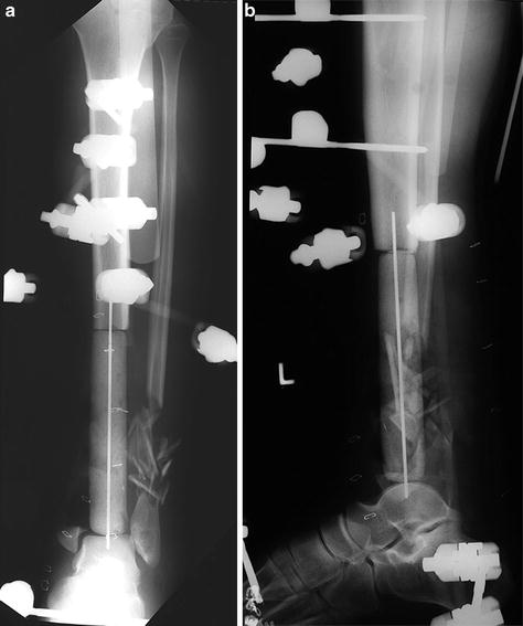
Fig. 2
(a) Anterior-posterior X-ray after debridement and placement of 13.5 cm spacer. The leg is stabilized with half pin distraction external fixator. The spacer was manufactured with 4.8 g of tobramycin, 2 g of vancomycin, and 40 g of bone cement. (b) Lateral X-ray. The foot is aligned in plantar neutral position. The antibiotic spacer is aligned by 0.0062 Steinman pin. The dome of the talus has to be drilled with a small-diameter drill bit to place the Steinman pin
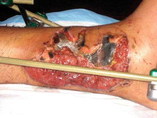
Fig. 3
Crush injury of the medial leg with loss of soft tissue requiring free flap. Granulation tissue with split skin graft is inadequate soft tissue coverage for spacer
3 Preoperative Problem List
1.
Loss of 13.5 cm of distal tibia including plafond
2.
Loss of soft tissue medial distal tibia requiring free flap
3.
Loss of posterior tibial artery with the foot surviving on anterior tibial artery and peroneal collateral arterioles
4.
Maintenance of plantar neutral position of the foot and alignment/flexibility of the toes during long reconstruction
4 Treatment Strategy
After multiple aggressive debridements, the reconstruction tunnel is commenced by placing the antibiotic spacer 26 mm in diameter. The extremity is maintained in good alignment with the half pin external fixator. The fixator is designed to give access to the plastic surgeons for the anastomosis of the flap. In this case an end-to-end hookup with the posterior tibial artery was accomplished. The flap was allowed to mature for 3 months before the bone reconstruction commenced. The flap is the most technical stage of the reconstruction. In sequential reconstruction, the soft tissues are resuscitated and healthy before starting the reconstruction of the tibia. The 3-month delay also creates a stable reconstruction tunnel with reactive membrane (Masquelet technique ) that will not collapse as the tibia is transported distally to arthrodesis under the flap. After transport of more than 1–2 months, the end of the transport will have developed a stable neocortex bone cap that requires docking site revision and squaring of the tibia shaft and dome of the talus to the bleeding bone.
5 Basic Principles
1.
The Ilizarov fixator proximal fixation block consists of a 5/8 ring connected to a full ring with 3 cm hexagonal sockets (an alternative is a 2/3 spatial open ring).
2.
The mid-tibia has a single-ring fixation block with three divergent 6 mm titanium hydroxyapatite pins.
3.
The distal fixation block is a single or double foot ring with opposed olive wires in the calcaneus and opposed olive wires in the talus. Small feet have a single ring with the talar wire on post. Large feet have two foot rings separated by 3 cm hexagonal sockets with the talar wires based on the proximal ring.
4.
 8: Femoral Bone Defect
 11: Bone Transport Over a Nail for Infected Tibial Nonunion and Bone Defect
 30: C3.3 Pilon Fracture Closed. Ilizarov Fixation with Limited Open Reduction of Joint Surface and Distal Tibia Bridging Distraction of Ankle Joint
 78: Two-Stage Treatment of a Chronic Foot Dislocation
 84: Lapidus Fusion with External Fixation
 65: Closed Correction of Club Foot with Ilizarov


Related posts:
8: Femoral Bone Defect
11: Bone Transport Over a Nail for Infected Tibial Nonunion and Bone Defect
30: C3.3 Pilon Fracture Closed. Ilizarov Fixation with Limited Open Reduction of Joint Surface and Distal Tibia Bridging Distraction of Ankle Joint
78: Two-Stage Treatment of a Chronic Foot Dislocation
84: Lapidus Fusion with External Fixation
65: Closed Correction of Club Foot with Ilizarov
Stay updated, free articles. Join our Telegram channel
Full access? Get Clinical Tree
