Fig. 1
A photograph showing significant shortening of her left lower extremity

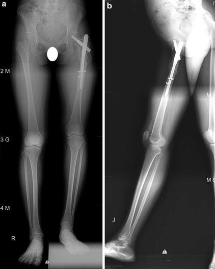
Fig. 2
(a) Long bone coronal and (b) sagittal views of her left lower extremity demonstrating a subtrochanteric fracture nonunion of the left femur with proximal intramedullary nail in place. Approximately 40 mm shortening and varus deformity at the fracture level (neck-shaft angle 119°) were found

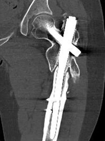
Fig. 3
Mid-coronal view from the computed tomography of her left femur demonstrating the nonunion at the fracture site
3 Preoperative Problem List
1.
Femoral shortening (4 cm), left
2.
Nonunion, subtrochanteric fracture, femur, left
3.
Varus deformity, subtrochanteric region, femur, left
4 Treatment Strategy
Nonunion should be managed with decortications, autogenous bone graft, and firm fixation with plating. Osteotomy should be placed below the nonunion site. About 4 cm gradual femoral lengthening with acute correction of varus deformity can be accomplished using the intramedullary lengthening device (PRECICE® , Ellipse, CA, USA) and multiple blocking screws.
5 Basic Principles
Placing osteotomy at the pre-injured site should be avoided because it can negatively affect successful bone regeneration. Firm fixation with augmentation plating and autogenous bone graft for the nonunion site and simultaneous lengthening using different levels of osteotomy is a good option for the treatment of nonisthmal femoral shaft nonunion.
6 Images During Treatment
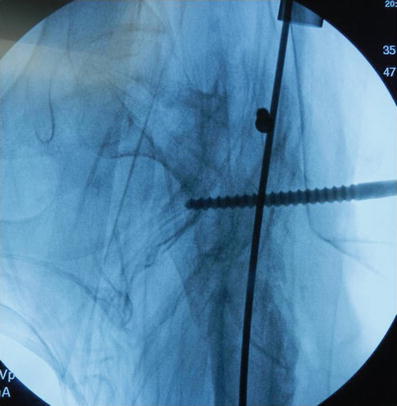
See Figs. 4, 5, 6, 7, and 8


