Figure 4.1
Herpes eruption on the interior portion of the left thigh
Clinical Differential Diagnosis
Drug eruption
Bullous pemphigoid
Pemphigus vulgaris
Linear IgA disease
Herpes simplex viral infection (type 2)
Bullous dermatophytosis
Bullous lichen planus
Histopathology
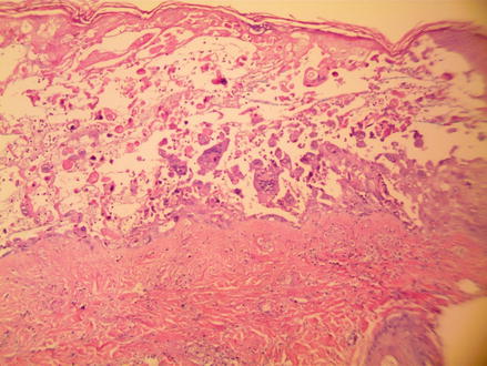
A shave biopsy was performed which measured 0.8 × 0.4 × 0.2 cm. Sections showed a fluid filled vesicle marked with extensive acantholysis and necrosis of the keratinocytes (Fig. 4.2) Scattered keratinocytes showed “nuclear molding” while other areas showed “nuclear margination” (Fig. 4.3).


