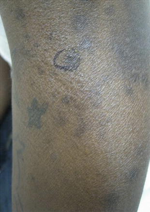
Figure 18.1
Syphilis, left lower arm

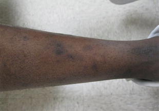
Figure 18.2
Syphilis, small dark, vesicular lesions on the median of the patient’s left leg

Figure 18.3
Syphilis, hyperpigmented patches found throughout the lower extremities
Clinical Differential Diagnosis
Drug eruption
Allergic dermatitis
Syphilis
Lichenoid dermatitis
Histopathology
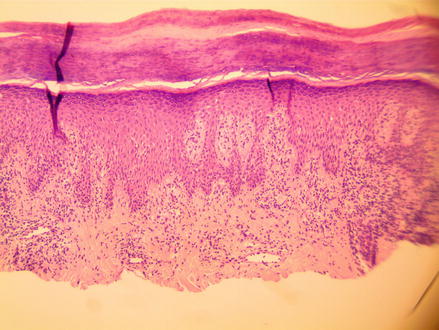
A shave biopsy was performed on the superior left arm (Fig. 18.1), and the medial left leg (Figs. 18.2 and 18.3) and both specimens measured about 0.6 × 0.5 × 0.1 cm. The low magnification showed a psoriasiform epidermal pattern with a chronic inflammatory infiltrate (Fig. 18.4). Sections showed a dense, interstitial infiltrate of lymphocytes and numerous plasma cells at high magnification (Fig. 18.5). An immunostain for spirochetes revealed numerous, short-rod shaped organisms in the lower epidermis and upper dermis (Fig. 18.6). Only at higher magnification, the corkscrew form of the spirochetes were appreciated (Fig. 18.7).


