Fig. 1
Clinical photograph of leg (a) and close-up photo of wound (b) 48 h after injury prior to second debridement and application of external fixator
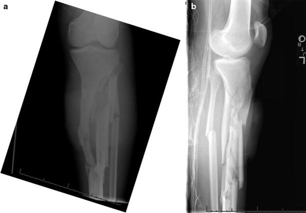
Fig. 2
AP (a) and lateral (b) X-rays of left leg showing comminuted segmental fractures
3 Preoperative Problem List
Polytrauma
Comminuted open segmental tibia fracture
Open wound with anterior soft tissue defect
4 Treatment Strategy
At the time of initial surgery, debridement was performed on both open tibia fractures, and the right was definitively treated with intramedullary rod. In the interest of minimizing operative time, a splint and provisional wound VAC were placed on the left. Forty-eight hours later after improvement in his clinical condition, repeat debridement was performed, and the leg was acutely shortened and angulated through the fracture to allow a tension-free primary closure of the wound. An incisional wound VAC was applied. The frame was extended distally and a corticotomy performed to allow concomitant distal lengthening after a week latency period.
The proximal frame remained statically locked in the angulated state for approximately 4 weeks to allow for primary wound healing; then, I began to slowly distract and correct the angular deformity. No bone grafting was performed at the fracture site. A removable dynamic splint was used to prevent ankle equinus. He was permitted weight bearing as tolerated, though this was complicated by the induced deformities.
5 Basic Principles
Adequate debridement of devitalized bone and soft tissue is the mainstay of treatment in high-energy open fractures . Treatment options included local rotational soft tissue defec t management with spanning external fixation followed by staged internal fixation with bone graft, as needed. However, after discussing my proposed treatment regimen, the family consented to the procedure herein described.
Another option would have been to correct the induced angular deformity and axial shortening at the proximal site. I opted to perform a distal corticotomy and lengthening to increase regional blood flow to the limb through corticotomy (Aronson 1994) and to shorten the total time in frame.
I used a removable anti-equinus orthotic during proximal deformity correction and distal distraction.
6 Images During Treatment
See Figs. 3, 4, 5, 6
 8: Femoral Bone Defect
 11: Bone Transport Over a Nail for Infected Tibial Nonunion and Bone Defect
 22: Bone Transport to a Knee Fusion and Secondary Intramedullary Nailing s/p Gunshot Wound
 78: Two-Stage Treatment of a Chronic Foot Dislocation
 84: Lapidus Fusion with External Fixation
 65: Closed Correction of Club Foot with Ilizarov


Related posts:
8: Femoral Bone Defect
11: Bone Transport Over a Nail for Infected Tibial Nonunion and Bone Defect
22: Bone Transport to a Knee Fusion and Secondary Intramedullary Nailing s/p Gunshot Wound
78: Two-Stage Treatment of a Chronic Foot Dislocation
84: Lapidus Fusion with External Fixation
65: Closed Correction of Club Foot with Ilizarov
Stay updated, free articles. Join our Telegram channel
Full access? Get Clinical Tree
