Figure 8.1
Condyloma acuminata on patient’s penile shaft
Clinical Differential Diagnosis
Epidermal nevus
Seborrheic keratosis
Condyloma acuminata
Condyloma lata
Histopathology
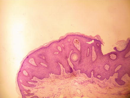
A shave biopsy was performed on the patient’s penile shaft. It measured 0.5 × 0.3 × 0.2 cm. Microscopic sections showed hyperplasia (thickened epithelium) and low papillomatosis that correlated to the cauliflower clinical appearance (Fig. 8.2). Some of the cells showed perinuclear cytoplasmic clearing (koilocytes) with an irregular nuclear contour (Fig. 8.3).


