Fig. 1
3D CT scans showing the extent of injury to the proximal tibial articular surface and metaphysis
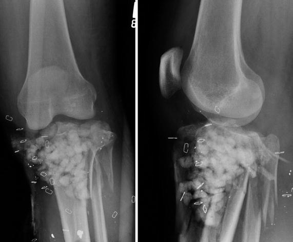
Fig. 2
AP (left) and lateral (right) images after initial debridement of injury with antibiotics beads in place
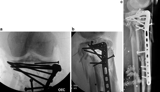
Fig. 3
(a–c) Intra-operative and post-operative radiographs following open reduction and internal fixation. AP (a), Lateral (b, c)
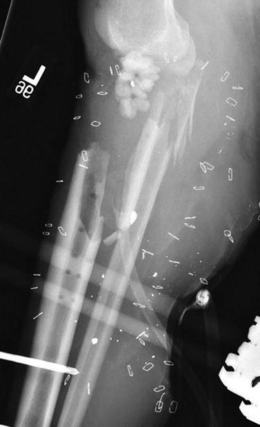
Fig. 4
Radiograph following extensive debridement of all infected hardware, bone and extensor mechanism. Bone loss is ~11 cm
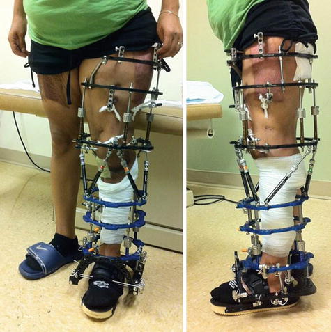
Fig. 5
Frontal (left) and lateral (right) clinical photographs showing frame construction. Note the use of standard “clickers” for lengthening and spatial frame struts in the middle of the frame for alignment
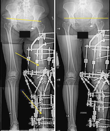
Fig. 6
Standing frontal alignment films showing frame construction and location of femoral and tibial regenerate bone (arrows) at the middle (left) and end of treatment (right)
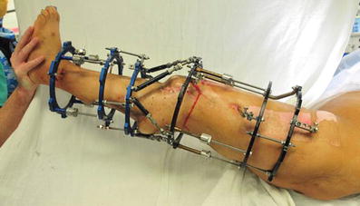
Fig. 7
Appearance of the leg and frame at the time of conversion to intramedullary fusion nail
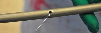
Fig. 8
Appearance of the 3 mm hole (arrow) to allow placement of a 2.7 locking screw at two locations in the nail to maintain compression of the knee fusion after IM nailing
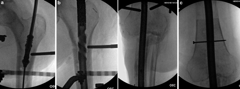
Fig. 9
(a–c) Intra-operative radiographs of placement of the intramedullary nail. (a) Reamer irrigator aspirator (RIA – Synthes USA) to prepare canal (b) Passage of the nail with the external fixation half pins in the unicortical position (c) 2.7 locking screw in place in the distal femur segment
3 Preoperative Problem List
1.
Deep infection (enterobacter and proteus).
2.
Loss of extensor mechanism.
3.
11 cm bone loss.
4.
Insulin dependent diabetes mellitus.
5.
Medial flap and skin graft.
4 Treatment Strategy
Stay updated, free articles. Join our Telegram channel

Full access? Get Clinical Tree


