Fig. 1
Pre-operative photos and radiographs: Left MAD = 26 mm medial; mMPTA = 85°, mMLTA = 106°, PPTA 63°, ADTA − 94°, Right MAD = 31 mm medial; mMPTA − 83°, mMLTA − 110°, PPTA 66°, ADTA − 100°
3 Preoperative Problem List
(a)
Hypertrophic nonunions of distal third in both tibial bones.
(b)
Complex six-component, three-planar deformity of right lower leg.
(c)
Complex five-component, two-planar deformity of left lower leg.
4 Treatment Strategy
According to the complexity of the deformities (severe angulation, torsion, shortening), existence of the consolidated fibulas and the condition of the soft tissues the plan is to perform a two-staged treatment: first stage – distal third osteotomy of both fibulas and gradual deformity correction using computer-assisted Ortho-SUV Frame. After deformity correction, as second stage, internal fixation by locking nails (closed, without opening the nonunion sites).
5 Basic Principles
(a)
Use of external fixation provides possibility of low-traumatic surgery, avoiding large incision and opening the bone fragments;
(b)
Use of computer-assisted Ortho-SUV Frame provides precise, one-step gradual deformity correction;
(c)
Deformity correction is to be made simultaneously at both lower legs ;
(d)
Reaming made during internal fixation stimulates the bone callus formation.
6 Images During Treatment
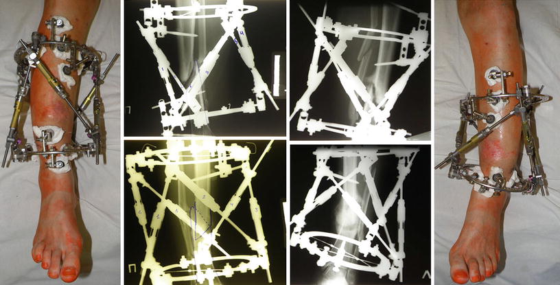
See Figs. 2, 3, 4, 5, 6, 7.


Fig. 2
The photos and radiographs after the surgery: Ortho-SUV Frames applied and osteotomies of fibulas were done

Fig. 3
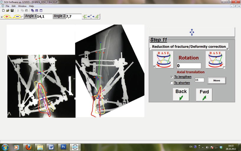
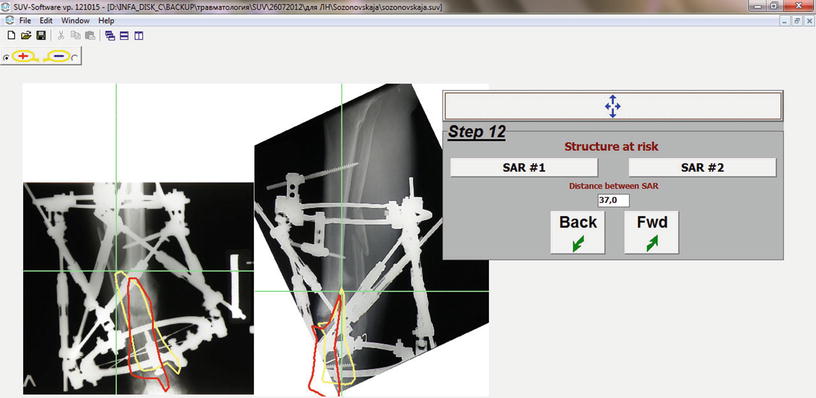
The Ortho-SUV software window at step 11: the deformities correction planning. The bone fragments anatomical axes were drawn. Yellow bone contour means initial mobile (corresponding) fragment position. Red bone contour shows created by software final position of mobile bone fragment


