Fig. 1
Pre-operative anteroposterior (a) and lateral (b) view radiographs (Copyright 2014, Rubin Institute for Advanced Orthopedics, Sinai Hospital of Baltimore)

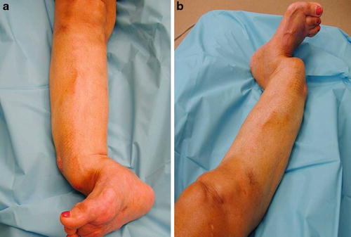
Fig. 2
Pre-operative anteroposterior (a) and lateral (b) view clinical photographs (Copyright 2014, Rubin Institute for Advanced Orthopedics, Sinai Hospital of Baltimore)

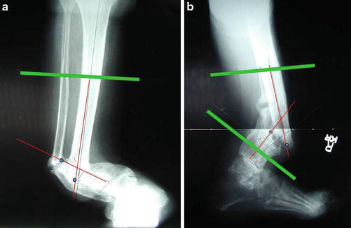
Fig. 3
Pre-operative anteroposterior (a) and lateral (b) view radiographs show deformity parameters (fracture method) (Copyright 2014, Rubin Institute for Advanced Orthopedics, Sinai Hospital of Baltimore)
3 Preoperative Problem List
Severe deformity
Long duration
Rheumatoid arthritis and diabetes mellitus
4 Treatment Strategy
Gradual deformity correction was combined with minimally invasive fixation. The foot was included when planning the deformity correction. Ankle range of motion was preserved, but the foot was included in the frame. The hypertrophic nonunion was distracted 0.5 mm/day at the concavity of the deformity. Additional bone grafting was not needed because the distraction allowed the regenerate process to occur. A cast was applied after frame removal to provide support and remained in place for 1 month.
5 Basic Principles
Gradually correct the deformity to avoid neurovascular injury. Also allow the hypertrophic nonunion to heal without bone graft. Address the ankle to avoid development of any equinus deformity. Include the foot in the frame for stability.


