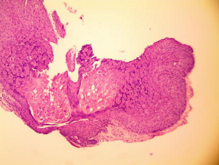
Figure 6.1
Molloscum contagiosum
Clinical Differential Diagnosis
Milia cyst
Pyogenic granuloma
Sebaceous hyperplasia
Molloscum contagiosum
Keratoacanthoma
Papular granuloma annulare
Fungal infections
Histopathology
The biopsy specimen from the left cheek measured 0.1 × 0.1 × 0.1 cm. Microscopic sections showed a lobular proliferation of keratinocytes. Some keratinocytes were enlarged and have a foamy vacuolated cytoplasm in the center of the lesion. Other keratinocytes were lost their nucleus and were brightly eosinophilic (Fig. 6.2).


