Procedure 13 In Situ Cubital Tunnel Decompression
 See Video 11: In Situ Cubital Tunnel Decompression
Indications
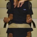
 In situ decompression of the ulnar nerve is indicated for patients with symptoms that are mild or intermittent (Table 13-1). If there is subluxation or instability of the ulnar nerve and/or ulnar nerve palsy owing to an abnormal osseous architecture of the elbow, in situ decompression is not indicated. In these situations, an anterior transposition is more appropriate to correct the anatomic problem.
Examination/Imaging
Clinical Examination


Surgical Anatomy
 The ulnar nerve is the terminal branch of the medial cord of the brachial plexus. Initially, the ulnar nerve lies medial to the axillary artery. In the upper arm, the ulnar nerve lies posteromedial to the brachial artery, posterior to the intermuscular septum, and anterior to the medial head of the triceps muscle. Approximately 8 cm proximal to the medial epicondyle is the arcade of Struthers, a thin fibrous band. At the elbow, the ulnar nerve travels posterior to the medial epicondyle and medial to the olecranon at the subcutaneous level, then it enters the cubital tunnel. The cubital tunnel is covered by fibroaponeurotic bands and the Osborne fascia (a ligament over the epicondylar groove), which is the fibroaponeurotic tissue between the two heads of the FCU. After passing through the cubital tunnel, the ulnar nerve travels deep into the forearm between the humeral and ulnar heads of the FCU. Distally, the ulnar nerve follows through the FCU to the deep flexor-pronator aponeurosis (Fig. 13-1).
Related posts:
Stay updated, free articles. Join our Telegram channel

Full access? Get Clinical Tree


