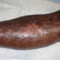
Fig. 1
Pre-operative clinical picture showing fistula (arrow) and atrophic changes over the skin
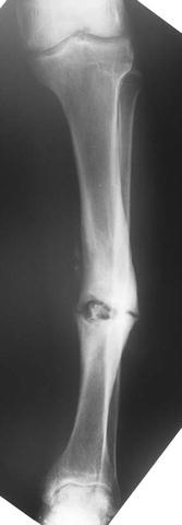
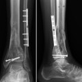
Fig. 2
Pre-operative AP X-ray demonstrates nonunion and varus deformity of a left tibia
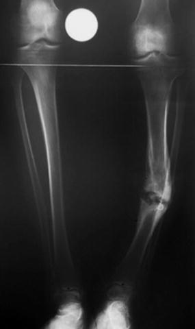
Fig. 3
Bilateral AP tibial X-ray demonstrating bone loss causing tibial shortening, nonunion, and deformity
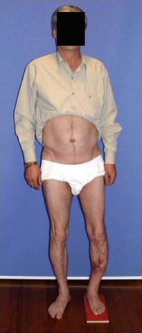
Fig. 4
Clinical picture of the patient standing. The tibia vara and shortening are apparent
3 Preoperative Problem List
4 Treatment Strategy
(a)
First session ( resection and debridement):
(i)
(ii)
Deep tissue samples are obtained for cultures, sensitivities, and Gram staining.
(iii)
(iv)
Six weeks of IV antibiotic therapy according to antibiogram (MRSA) results. The CRP levels should be checked to confirm elimination of the infection.
(b)
Second session(bone transport over IM nail ):
(i)
As a second stage the beads and the cement rod are replaced with an antegrade intramedullary nail, which is locked proximally. A circular external fixator (Ilizarov type) consisted of three rings is applied, and a corticotomy is performed at the proximal tibia (arrow) for lengthening and transport (Fig. 7a, b). Each ring is fixed to the corresponding tibial segment.
(c)
Get Clinical Tree app for offline access
Third session (ex. fix. removal and IM nail locking):
(i)
 8: Femoral Bone Defect
 28: Proximal Tibial Bone Defect Treated with Intentional Deformity and Bone Transport
 30: C3.3 Pilon Fracture Closed. Ilizarov Fixation with Limited Open Reduction of Joint Surface and Distal Tibia Bridging Distraction of Ankle Joint
 78: Two-Stage Treatment of a Chronic Foot Dislocation
 84: Lapidus Fusion with External Fixation
 65: Closed Correction of Club Foot with Ilizarov


Related posts:
8: Femoral Bone Defect
28: Proximal Tibial Bone Defect Treated with Intentional Deformity and Bone Transport
30: C3.3 Pilon Fracture Closed. Ilizarov Fixation with Limited Open Reduction of Joint Surface and Distal Tibia Bridging Distraction of Ankle Joint
78: Two-Stage Treatment of a Chronic Foot Dislocation
84: Lapidus Fusion with External Fixation
65: Closed Correction of Club Foot with Ilizarov
Stay updated, free articles. Join our Telegram channel
Full access? Get Clinical Tree
