Procedure 10 Extensor Tendon Repair in Zones 1 to 5
 See Video 7: Turnover Tendon Flap for PIP Extensor Tendon Injury


Surgical Anatomy
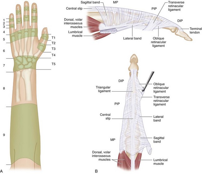
 Extensor tendon injuries are divided into nine zones for the fingers and seven zones for the thumb numbered from distal to proximal (Fig. 10-2A). An easy way to remember these zones is to bear in mind that the odd-numbered zones are located over the joints. Therefore zones 1, 3, 5, and 7 are located over the distal interphalangeal (DIP) joint, proximal interphalangeal (PIP) joint, metacarpophalangeal (MCP) joint, and wrist, respectively.
 The extensor tendon is thick over the MCP joint but becomes broad and thin over the dorsum of the proximal phalanx, where it divides into three portions. The central portion becomes the central slip that inserts into the base of the middle phalanx, and the lateral portions join the lateral bands. The tendons of the lumbrical and interosseous muscles form the lateral bands. These lateral bands come together over the middle phalanx and form the most distal part of the extensor tendon that inserts into the base of the distal phalanx (Fig. 10-2B).


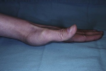
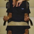
 Lacerations to the extensor pollicis longus (EPL) tendon cannot be tested by simply examining extension of the thumb interphalangeal (IP) joint. The IP joint may be extended by the extensor pollicis brevis (EPB) and the thenar intrinsic muscles via their insertions on the extensor expansion. The EPL is tested by placing the patient’s hand flat on a table and asking the patient to lift the thumb off the table (Fig. 10-3).
Positioning
 Extensor tendon repairs can be done under a digital block (zones 1, 2, and 3) or a wrist block (zones 4 and 5). The use of a forearm tourniquet (placed distally over the nonmuscular portion of the forearm) will allow the patient to tolerate the tourniquet for a longer period (30 to 40 minutes) compared with the usual placement over the arm (20 to 30 minutes). The patient is positioned supine with the affected extremity on a hand table.
Exposures
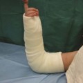
 The existing lacerations are incorporated to raise wide flaps with a broad tip.
 Full-thickness skin flaps are raised superficial to the paratenon.
Zone 1 Injury
Clinical Examination
 Patients present with a flexed posture of the DIP joint (mallet finger) and inability to actively extend the DIP joint (Fig. 10-4).

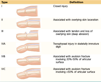
 Doyle has classified mallet finger into four types (Table 10-1).
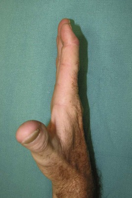
Table 10-1 Classification of Mallet Finger
Procedure
Step 1
 The proximal end of the tendon is mobilized for 5 to 8 mm such that the tendon ends lie close to each other with the joint in neutral position.
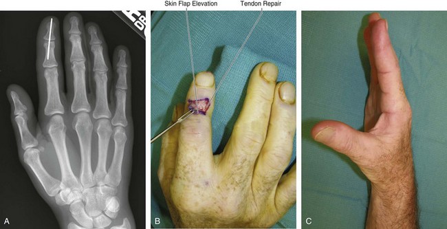
Step 2
 The DIP joint is pinned in extension or slight hyperextension using a 0.045-inch K-wire (Fig. 10-5A).
Step 3
Related posts:
 28: Pronator Teres Rerouting
 33: Correction of Swan-Neck Deformity in the Rheumatoid Hand
 89: Total Wrist Fusion
Stay updated, free articles. Join our Telegram channel

Full access? Get Clinical Tree


